视网膜屏幕_视网膜脱离-了解这一点很重要
视网膜屏幕
The human eye can be compared with the device of the camera, the lens of which is the cornea with the lens, and the film is the retina, an extremely complex multi-layer structure that is connected to the visual divisions of the brain with the help of nerve fibers. Therefore, we can assume that the retina is a part of the brain.
可以将人眼与摄像头的设备进行比较,摄像头的透镜是角膜和透镜,而胶片是视网膜,这是一种极其复杂的多层结构,通过眼睛与大脑的视觉区域相连。帮助神经纤维。 因此,我们可以假设视网膜是大脑的一部分。
Retinal detachment most often takes the patient by surprise — before it appears, a person may have excellent vision and may not present any complaints. The speed of propagation of the process is quite rapid, the treatment in the majority of cases is surgical.
视网膜脱离通常会使患者感到惊讶-在出现之前,一个人可能具有良好的视力,可能不会出现任何不适。 该过程的传播速度相当快,大多数情况下的治疗是外科手术。

The timeliness of the operation gives a chance to preserve vision; in Germany, according to the standard, the operation must be performed within 24 hours after the diagnosis. There are no such standards in Russia. But for each patient, I say that retinal detachment is “like fresh frozen fish” — in a couple of days it’s already “not the first freshness”.
手术的及时性为维护视觉提供了机会。 在德国,根据标准,必须在诊断后24小时内进行操作。 俄罗斯没有此类标准。 但是对于每位患者,我说的是视网膜脱离“就像新鲜的冷冻鱼一样”-在几天之内,它已经不是“第一次新鲜”。
There are many methods of treatment for retinal detachment, they differ in the mechanism of action, they can be combined with each other, there are no better or worse ones among them — all are very individual.
视网膜脱离的治疗方法有很多,它们的作用机理不同,可以相互结合,其中没有好坏之分-都是非常个别的。
Prevention (but not immunity) for retinal detachment exists — it is laser coagulation of areas on the retina, which may be the cause. These are certain types of dystrophies, tractions, breaks — but, unfortunately, not every patient believes in the need for these procedures, especially if nothing is bothering you.
存在视网膜脱离的预防措施(但不是免疫措施)-视网膜区域的激光凝结可能是原因。 这些是某些类型的营养不良,牵引,休息-但是,不幸的是,并非每个患者都认为需要这些程序,尤其是在没有任何事情困扰您的情况下。
视网膜断开的原因 (Causes of retina disconnect)
Detachment is the separation of rods and cones, we call them neuroepithelium, from the underlying pigment epithelium by the accumulation of fluid between them. This disrupts the power of the outer layers of the retina, which leads to rapid loss of vision.
分离是棒和视锥的分离,我们称它们为神经上皮,是通过它们之间的积聚而从下面的色素上皮中分离出来的。 这会破坏视网膜外层的力量,从而导致视力Swift丧失。
The possibility of detachment due to the characteristics of the structure of the retina, I wrote about this in previous posts.
由于视网膜结构的特征,有可能发生脱离,我在之前的文章中对此进行了介绍。

Retinal detachment in its type may be dystrophic (rhematogenous), traumatic and secondary. The secondary is not considered as an independent clinical form, but is only a complication of the underlying eye disease — inflammation, tumor, vascular or congenital diseases.
视网膜脱离的类型可能是营养不良(生发性),外伤性和继发性的。 继发性疾病不被认为是独立的临床形式,而仅仅是潜在的眼部疾病(炎症,肿瘤,血管或先天性疾病)的并发症。
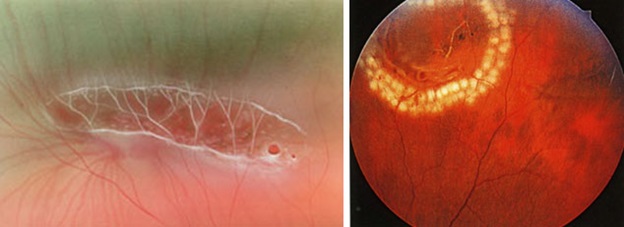
The reason for regmatogenous (regma — rupture) retinal detachment, or, they say, primary detachment, as is already clear, is a rupture or rupture of the retina. As a rule, the gap occurs somewhere on the periphery, close to the equator of the eye, in the area of thinning and dystrophy.
他们已经说过,视网膜变性或视网膜原发脱离的原因是视网膜形成或破裂。 通常,缝隙发生在变薄和营养不良的区域中,靠近眼睛的赤道的某个地方。

Types of dystrophies, dangerous in terms of delamination, have already been mentioned in posts earlier:
较早前的帖子中已经提到了营养不良的类型,它们在分层方面很危险:
How to “sew up” the retina and should it be done?如何“缝合”视网膜,应该怎么做?
The names of dystrophies are non-standard: “Lattice dystrophy”, “dystrophy by the type of snail track”, “traction”, “frivolous”, “white without impression”, “perforated breaks with and without lid”, “valve breaks” and others.
营养不良的名称是非标准的:“晶状体营养不良”,“按蜗牛轨道类型的营养不良”,“牵引力”,“轻浮”,“白色无印痕”,“有盖和无盖的穿Kong破裂”,“瓣膜破裂” “ 和别的。
This is how dystrophic foci require coagulation (before and after the procedure).这就是营养不良灶需要凝固的方式(手术前后)。
预防视网膜脱离 (Prevention of retina detachment)
From prophylaxis, the onset is intentional, because a timely preventive procedure can reduce the risk of exfoliation by an order of magnitude.
从预防上来说,起病是有意的,因为及时的预防措施可以使剥落的风险降低一个数量级。
Since the previous posts described in detail how this happens, I want to draw attention to some points.
由于前面的文章详细介绍了这种情况,因此,我想提请注意几点。
1. We say that, as a rule, laser coagulation is not a painful procedure, but there is always an amendment to individual sensitivity. In some cases, it can be painful, and in particularly sensitive ones it can even be very painful. The following factors play a role:
1.我们说,激光凝结通常不是一个痛苦的过程,但是对个人敏感性总是有修正的。 在某些情况下,它可能很痛苦,在特别敏感的情况下,甚至可能非常痛苦。 以下因素起作用:
— coagulation volume and localization of zones,
—凝血量和区域定位,
— laser type and model,
—激光类型和型号,
— patient's fit and behavior during it,
—在此期间患者的健康状况和行为,
— palpebral anatomy (“deep-set” eye, large nose, etc.)
—睑裂解剖(“深陷”的眼睛,大鼻子等)
— Experience a laser surgeon and the right choice of contact lenses.
—体验激光外科医生和正确选择隐形眼镜的经验。
Conclusion: if you have a high pain threshold, you are afraid or you are uncomfortable during the procedure — be sure to warn the doctor in advance or during it, with the help of drugs we can greatly facilitate the process of laser coagulation.
结论:如果您的疼痛阈值高,在手术过程中会感到害怕或不舒服-请务必提前或在手术过程中警告医生,借助药物,我们可以大大促进激光凝固的过程。
胎盘疾病的诊断 (Diagnostics of retardium diseases)
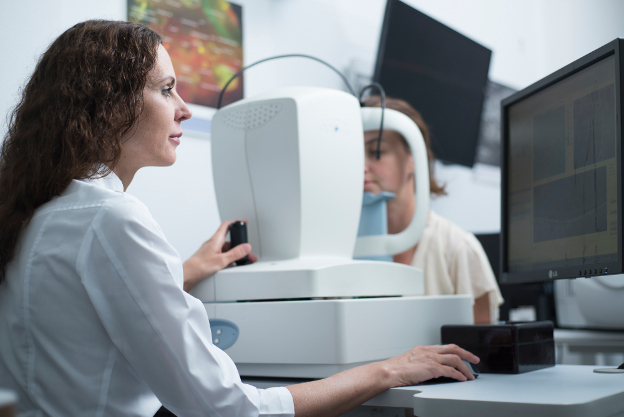
The process of optical coherent tomography of the retina (OCT) in our clinic.我们诊所的视网膜光学相干断层扫描(OCT)过程。
Diagnosis of retinal detachment involves primarily a complete ophthalmologic examination with eye examination, intraocular pressure, fundus examinations in various ways — contact and non-contact. You may also need to inspect both vertically and horizontally.
视网膜脱离的诊断主要包括对眼科的全面检查,包括眼部检查,眼内压,眼底检查等多种方式-接触式和非接触式。 您可能还需要垂直和水平检查。
Special additional diagnostic methods are:
特殊的附加诊断方法是:
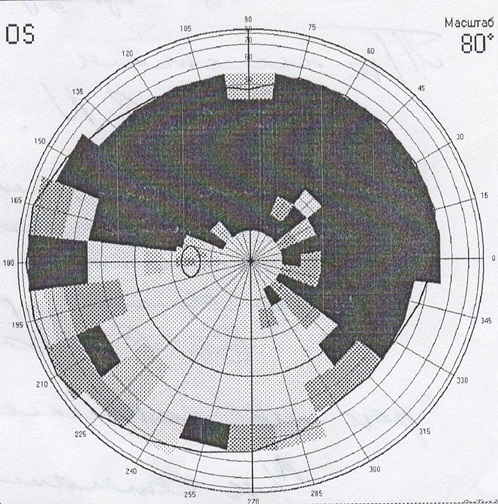
1. Perimetry. A frequent symptom of detachment is «veil» or «curtain» in front of the eye, it looks like this:
1.视野测定法。 眼球脱离的常见症状是眼前的“面纱”或“窗帘”,看起来像这样:
2. Ultrasound scanning in 2-D or 3-D mode. Allows you to define a detachment through the opaque optical medium of the eye or areas inaccessible to the examination… You can determine its height, the contents under it, the relief, the thickness of the shells — this is important.
2.在2-D或3-D模式下进行超声扫描。 允许您定义不透明的眼睛光学介质或检查所无法触及的区域的分离……您可以确定其高度,其下的内容物,浮雕和壳的厚度-这很重要。

3. Electrophysiology — as an eye cardiogram — captures electrical potentials from functioning areas of the retina, indicating the extent of its damage.
3.电生理学–作为眼动心电图–捕获视网膜功能区域的电势,表明其受损程度。
4. Optical coherent tomography — we obtain linear retinal sections to determine the anatomical parameters of the smallest sections in the central and close to it areas. The result may be a two- or three-dimensional image.
4.光学相干断层扫描-我们获得线性视网膜切片,以确定中央区域及其附近区域中最小切片的解剖学参数。 结果可以是二维或三维图像。
5. X-ray CT of the retina — provides visualization of the structures of the eye, down to the smallest details.
5.视网膜的X射线CT-可视化眼睛结构,直至最小的细节。
6. MRI — shows the degree of violation and makes it possible to create a three-dimensional image of the eye.
6. MRI-显示侵犯的程度,并可以创建眼睛的三维图像。
视网膜脱离的症状 (Symptoms of the retina detachment)
Comparing the eye with a camera in which there is a film, we can say that somewhere on the edge of the frame there appeared a scratch of the emulsion layer. Well, what of this, you say, because almost the entire frame and the most important thing — the center of the “composition” — is still visible well. It turns out that this is not true. Through the gap begins to penetrate the fluid, flowing under the retina and thereby peeling it from the underlying choroid. On film, it looks as if an emulsion layer starts to blister around the scratch and peel off from the substrate. A person at this moment sees a rather characteristic picture of a “gray curtain” at the edge of the visual field. Depending on the location of the gap, the “curtain” can either spread quickly (within several tens of hours), closing the entire field of view, or crawl more smoothly (for weeks)
将眼睛与装有胶卷的相机进行比较,可以说在镜框边缘的某个地方出现了乳剂层的划痕。 好吧,这就是您所说的,因为几乎整个框架和最重要的部分(“构图”的中心)仍然清晰可见。 事实证明这是不正确的。 穿过缝隙开始渗透流体,在视网膜下方流动,从而将其从下面的脉络膜剥离。 在胶片上,似乎乳液层开始在划痕周围起泡并从基材上剥离。 此时,一个人在视野的边缘看到了相当有特色的“灰色窗帘”图片。 取决于间隙的位置,“窗帘”可以快速传播(在几十小时内),关闭整个视场,或者可以更平滑地爬行(持续数周)

Vision in the development of retinal detachment视网膜脱离发展的愿景
Quite typical of fresh retinal detachment is a symptom of “morning improvement”, when a person in the morning (after a long inactive lying position) reveals a significant improvement (reduction of the curtain, its blanching and the ability to see through it). By lunchtime it gets worse again, and by evening it gets worse.
新鲜的视网膜脱离典型表现为“早晨改善”的症状,当一个人在早晨(长时间处于不活动的卧姿后)表现出明显的改善(窗帘的减少,其变白和能够透视的能力)。 到午餐时间又变得更糟,到晚上变得更糟。
It is clear that if the gap is located in the upper parts of the eye, the fluid quickly goes down and the exfoliation passes rapidly. If the gap is located below, then the detachment slowly «crawls» up, and the progression will be slower. However, the adhesions between the retinal zones and scars will be more pronounced — the time for their formation will be more.
显然,如果间隙位于眼睛的上部,则液体会Swift下降,并且剥落会Swift通过。 如果间隙位于下方,则分离会缓慢地“爬行”,并且进展会变慢。 但是,视网膜区域和疤痕之间的粘连会更加明显-形成它们的时间会更长。
治疗 (Treatment)
Treatment is necessary, and only surgical, there is no other way out. No drops, ointments, tablets, injections, absorbable means do not help, but only take time, which allows the detachment to develop further and further. The sooner competent surgical treatment is carried out, the better results it gives and the more it is possible to restore sight. The goal of surgical treatment was formulated more than 100 years ago and is to close (block) the retinal break.
治疗是必要的,只有手术,没有其他出路。 没有滴剂,软膏剂,片剂,注射剂,可吸收手段无济于事,而只需要时间,这使得分离越来越深。 进行胜任的外科手术治疗越早,其效果越好,恢复视力的可能性就越大。 手术治疗的目标是在100多年前制定的,目的是闭合(阻塞)视网膜裂Kong。
At the initial stage of the disease, there is usually no need to enter the eye, and surgery consists of a local external impression in the projection of the rupture. To do this, use special seals made of soft silicone, which press the area of the gap, thus blocking it.
在疾病的初始阶段,通常不需要进入眼睛,而手术包括在破裂部位投射出局部的外在印象。 为此,请使用由软硅胶制成的特殊密封垫,该密封垫会压住间隙区域,从而阻塞间隙。

The scheme of episcleralisation巩膜化方案
As soon as the opening in the retina closes, everything miraculously gets better, the “curtain” disappears, the vision begins to recover. Peripheral vision is restored first, the person discovers that the “review” is almost normal, and later on it really becomes normal. The periphery of the retina is fairly stable, and as soon as it becomes in its anatomical place, it immediately begins to “work” and recovers well even with long periods of retinal detachment. With central vision, things are not so simple. The most favorable cases are when the detachment did not have time to “crawl” to the center. For example, if the vision in the center remained 1.0, and the “curtain” closed the half of the field of view, after a successful operation, the vision could recover to 1.0, and the curtain would disappear.
视网膜开口一旦关闭,一切都会奇迹般地变得更好,“窗帘”消失,视力开始恢复。 首先恢复周围的视力,此人发现“检查”几乎是正常的,然后才真正恢复正常。 视网膜的外围相当稳定,一旦进入解剖位置,它就会立即开始“工作”,即使视网膜脱离很长时间也能很好地恢复。 有了中心视野,事情就不会那么简单了。 最有利的情况是分队没有时间“爬行”到中心。 例如,如果中心的视力保持为1.0,并且“窗帘”关闭了视场的一半,则在成功操作之后,视力可能会恢复到1.0,并且窗帘会消失。
If the detachment managed to close the central zone, after a successful operation, the central vision, unfortunately, can no longer fully recover. What will be the visual acuity after surgery in this case depends on a number of factors. The most important of them are the time during which the central zone of the retina has exfoliated, and the state of the blood supply to the retina, which directly depends on the age and degree of myopia (if there is one).
如果该分队成功地关闭了中央区域,则在成功手术后,不幸的是,中央视野将无法完全恢复。 在这种情况下,手术后的视敏度取决于许多因素。 其中最重要的是视网膜中央区域脱落的时间,以及视网膜血液供应的状态,这直接取决于近视的年龄和程度(如果有)。
Restoration of central vision occurs slowly and usually almost ends at 3 months. Further improvement may continue, but at a still slower pace, and we observe that after a year, and after 3 years, visual acuity improves a little.
恢复中枢视力的过程很缓慢,通常在3个月时就结束。 进一步的改善可能会继续,但速度仍然会变慢,我们观察到一年后和三年后,视敏度会有所改善。

Changes in the structure of the retina after detachment, worsening the “picture”脱离后视网膜结构的变化,使“图像”恶化
If a person with retinal detachment is not operated on time or operated unsuccessfully, then the detachment remains and continues to develop, moreover, the so-called “proliferative process” begins in the vitreous.
如果患有视网膜脱离的人未按时手术或手术不成功,则该脱离仍然存在并继续发展,此外,所谓的“增生过程”从玻璃体开始。
The eye, as you know, has the shape of a sphere, and we already know that it has a lens, a retina film, besides this, inside the eye is filled with liquids. These fluids are almost 98-99% water, but with very substantial additives. The anterior part of the eye is bounded by the cornea on one side and the iris-lens block on the other. This part of the eye is more responsible for optics and is filled with anterior chamber intraocular fluid. In terms of its properties and appearance, it hardly differs from simple water with the addition of a complex set of minerals and salts.
如您所知,眼睛具有球体的形状,而且我们已经知道它具有晶状体,即视网膜膜,此外,眼睛内部充满了液体。 这些流体几乎是98-99%的水,但含有非常大量的添加剂。 眼睛的前部在一侧被角膜所包围,而在另一侧则被虹膜透镜所阻挡。 眼睛的这一部分负责光学,并充满前房眼内液。 就其性质和外观而言,它与简单的水几乎没有什么不同,只是添加了一组复杂的矿物质和盐。
Another thing is the fluid in the posterior part, bounded by the lens, ciliary body and retina. This liquid is called the vitreous body, it has the consistency and the appearance of a gel or frozen jelly. In addition, the basis of the vitreous body is a framework in the form of a voluminous lattice of collagen fibers.
另一件事是后部的液体,以晶状体,睫状体和视网膜为界。 这种液体称为玻璃体,具有凝胶状或冻冻状的稠度和外观。 另外,玻璃体的基础是胶原纤维的大量格子形式的框架。
When retinal detachment the vitreous body never remains indifferent. In the initial period, only small violations of its structure are observed, manifested in the form of various inclusions floating in the field of view. With a long-existing detachment in the framework of the vitreous body, strands develop, which, like ropes, attach to the retinal surface and, slowly contracting, retract the retina to the center of the eyeball. This process is called vitreoretinal proliferation, which ultimately leads to the formation of the so-called «funnel» retinal detachment. In such a situation, reconstructive surgery is required, in terms of the quality of a much higher level. Close this gap seals is almost impossible, and not enough. The main task is to clean the surface of the retina from the strands of the vitreous body, straightening it.
当视网膜脱离时,玻璃体永远保持冷漠。 在最初阶段,仅观察到其结构的一些小违规,表现为视野中漂浮的各种夹杂物。 随着玻璃体框架内长期存在的脱离,会形成股线,就像绳索一样,股线会附着在视网膜表面,然后缓慢收缩,使视网膜缩回眼球中心。 该过程称为玻璃体视网膜增生,其最终导致形成所谓的“漏斗”视网膜脱离。 在这种情况下,就更高水平的质量而言,需要进行重建手术。 封闭这种间隙密封几乎是不可能的,而且还远远不够。 主要任务是清洁玻璃体股线的视网膜表面,使其变直。
A picture of a severe form of proliferative retinopathy严重形式的增生性视网膜病的照片
For this purpose, special methods of so-called vitreoretinal surgery are used. Its essence lies in the fact that through point punctures with long and thin instruments the surgeon enters the eye and removes cords, freeing the retina and straightening it. The process itself is very similar to the painstaking work of the master, who collects a model of a XVIII century sailboat inside the bottle through the neck of the bottle with long tweezers and scissors.
为此,使用所谓的玻璃体视网膜手术的特殊方法。 其本质在于以下事实:外科医生通过细长的器械进行点刺穿刺,使外科医生进入眼内并去除了脐带,从而使视网膜游离并变直。 这个过程本身与船长的艰苦工作非常相似,船长用长镊子和剪刀通过瓶子的颈部收集了十八世纪帆船的模型。

Vitrectomy (schematically)玻璃体切除术(示意性地)
This operation is very thin and complex, if you remember that the retina is extremely delicate and fragile nerve tissue, and almost every part of it is responsible for any part of the vision. During the operation, the doctor looks inside the eye through his anterior segment, peeks «through the pupil.» This requires a high transparency of the optical media, that is, the cornea lens and lens should be as transparent as possible. If the lens is cloudy, that is, the patient has a cataract, then, as a rule, at the initial stage, the lens is replaced with an artificial one, and only then proceed to «repair» the retina.
如果您还记得视网膜是非常脆弱的神经组织,那么该操作非常薄且复杂,几乎每个部分都对视力的任何部分负责。 在手术过程中,医生通过他的前眼向内看,“通过瞳Kong”窥视。 这要求光学介质具有高度透明性,即角膜透镜和透镜应尽可能透明。 如果晶状体浑浊,即患者患有白内障,那么通常在初始阶段,将晶状体替换为人工晶状体,然后才开始“修复”视网膜。
How we do this is discussed in these posts:
这些帖子中讨论了我们如何做到这一点:
Cataract: it is waiting for you personally (if you live, of course)白内障:它正在亲自等你(当然,如果你活着的话) We implant an artificial lens (you will need it after 60 years)我们植入一个人工晶状体(60年后您将需要它)
In addition, the natural lens, due to its anatomical location, often interferes with the work on the peripheral parts of the retina. In these cases, it is also necessary to change the lens to an artificial one, otherwise the untreated areas of the peripheral retina may not allow its anatomical fit to be achieved.
另外,天然晶状体由于其解剖学位置,常常会干扰视网膜外围部分的工作。 在这些情况下,也有必要将晶状体更换为人造晶状体,否则周边视网膜的未治疗区域可能无法实现其解剖结构。
The operation is performed in the “dark room”, that is, only the light guide in the surgeon’s hand or the additional light source “chandelier” lights up the working field as a chandelier, the light of the microscope is turned off.
该操作在“暗室”中进行,即,只有手术医生手中的光导或附加光源“枝形吊灯”作为枝形吊灯照亮工作区域,显微镜的光才会关闭。
Outwardly, it happens like this (photo from the operating room)从外面看,是这样的(手术室的照片)
After complete purification of the retinal surface from the vitreous cords, it must be straightened and placed on the choroid, that is, get its anatomically correct position inside the eye. For these purposes, the so-called “heavy water” is often used — liquid perfluororganic compound (PFOS). By its properties, this substance hardly differs from ordinary water, but due to its higher molecular weight it acts as a press on the surface of the retina, smoothing and pressing it. “Heavy water” copes very well with the detachment, in addition, it is absolutely transparent, and the eye filled with this liquid begins to see almost immediately. The main disadvantage is that the eye does not tolerate it for a long time. A maximum of a couple of weeks, but in practice for more than 7-10 days, it is undesirable to leave this liquid in the eye.
从玻璃体细胞中完全纯化视网膜表面后,必须将其拉直并放在脉络膜上,即在解剖学上使其位于眼睛内部的正确位置。 为了这些目的,通常使用所谓的“重水”-液态全氟有机化合物(PFOS)。 由于其性质,该物质与普通水几乎没有区别,但是由于其较高的分子量,它可以充当视网膜表面的挤压物,使其平滑并挤压。 “重水”可以很好地解决这种分离,此外,它绝对透明,充满这种液体的眼睛几乎可以立即看到。 主要缺点是眼睛长时间无法忍受。 最多需要几个星期,但实际上要持续7-10天以上,因此不宜将这种液体留在眼中。
视频葡萄膜外科 (VIDEO VITRECTOMY SURGERY)
Unfortunately, retinal glue has not yet been invented, but the laser turned out to be very effective. The laser “welds” the retina to the underlying tissues along the edges of all the breaks. After the application of laser coagulates, local inflammation occurs, and then a microtubule is gradually formed (5-7 days) on the choroid. Therefore, it makes sense to leave “heavy water” in the eye for a week. In some cases, this is enough to keep the retina in place, but it may be necessary to continue to hold the retina to form more durable adhesions. In such cases, silicone oil is used, which fill the eye cavity.
不幸的是,视网膜胶尚未被发明,但是激光被证明是非常有效的。 激光沿着所有断裂的边缘将视网膜“焊接”到下面的组织。 施加激光凝结后,发生局部炎症,然后在脉络膜上逐渐形成微管(5-7天)。 因此,将“重水”放在眼中一周是有意义的。 在某些情况下,这足以将视网膜保持在适当的位置,但是可能有必要继续保持视网膜以形成更持久的粘连。 在这种情况下,将使用硅油填充眼Kong。
Variants of silicone oil of varying degrees of viscosity各种粘度的硅油变体
Silicone is a clear, viscous liquid, the tissues almost do not react to it, so you can leave it in the eye for much longer. Silicone is not so good straightens and presses the retina, but to keep the retina unfolded, it fits perfectly.
硅酮是一种透明的粘稠液体,组织几乎不会对其产生React,因此您可以将其留在眼中更长的时间。 硅胶不是很好拉直并按压视网膜,但要使视网膜保持展开状态,它会非常适合。
The eye filled with silicone almost immediately begins to see, the retina retains its anatomical position, its functions are restored, and the adhesions in the areas of laser coagulates become very durable over time. One of the features of silicone is a change in the optical characteristics of the eye to the plus side by 4–5 diopters. Usually silicone is in the eye for about 2-3 months, after which the retina no longer needs any “props” and can be safely removed. This is also an operation, but not as complicated and voluminous as the previous ones.
充满硅酮的眼睛几乎立即开始可见,视网膜保持其解剖位置,恢复其功能,并且随着时间的流逝,激光凝结区域的粘连变得非常持久。 硅树脂的特征之一是眼睛的光学特性向正侧变化了4–5屈光度。 通常,硅树脂会在眼睛中停留约2-3个月,此后,视网膜不再需要任何“道具”,并且可以安全地将其去除。 这也是一项操作,但不如先前的操作那么繁琐。
In some cases, changes in the internal eye structures are so pronounced that the only option today is to have at least residual vision, or to keep the eye as an organ — is the constant presence of silicone in the eye cavity. In these cases, silicone can remain in the eye for many years, even decades.
在某些情况下,内部眼部结构的变化如此明显,以至于当今唯一的选择是至少具有残留的视力,或保持眼睛为器官-眼腔中不断存在有机硅。 在这些情况下,有机硅可以在眼睛中保留很多年,甚至数十年。
In addition to «heavy water» or silicone oil, for the same purpose, various gases or air are sometimes used. Principle one, from the inside, press the retina for a while with the air bubble until the scars get stronger. Any gas, especially air, dissolves over time into the eye fluid and disappears. The air dissolves within 1-2 weeks, the gas can be in the eye for up to 2 months. Unlike silicone, a person with injected gas sees practically nothing except light and bright objects. Gradually, the boundary appears between the gas bubble and the ophthalmic fluid. The patient notes the fluctuations of the bubble when moving the head. As the gas is absorbed from above, the image begins to open and, eventually, the entire field of vision becomes clear. Thus, surgical treatment takes place in one stage — you do not need to remove air or gas.
除了“重水”或硅油以外,出于相同的目的,有时还会使用各种气体或空气。 原理一,从内部用气泡按压视网膜一会儿,直到疤痕变强。 任何气体,特别是空气,随着时间的推移会溶解到眼液中并消失。 空气在1-2周内溶解,气体可以在眼中长达2个月。 与硅树脂不同,注有气体的人几乎看不到任何东西,只有明亮的物体。 逐渐地,边界出现在气泡和眼科液体之间。 患者在移动头部时会注意到气泡的波动。 随着气体从上方被吸收,图像开始打开,最终整个视野变得清晰。 因此,外科手术治疗分一个阶段进行-您无需去除空气或气体。
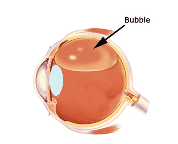
This is how gas looks schematically, in the stage of resorption in the vitreous cavity.这是在玻璃体腔内吸收阶段中气体的示意图。
All methods and substances used today in vitreal surgery are only tools for one big task — restoring vision after retinal detachment. Each case of detachment is individual and only the surgeon can decide what is best for a particular eye and for a particular patient. We can say with confidence that, using and combining modern methods, we manage to cope with almost any detachment. Another question is how damaged they are, how long the nerve cells of the retina did not work and to what extent they can recover after receiving its complete anatomical fit.
如今,玻璃体手术中使用的所有方法和物质仅是一项重要任务的工具-视网膜脱离后恢复视力。 每种脱离情况都是个别的,只有外科医生才能决定哪种方法最适合特定的眼睛和特定的患者。 我们可以充满信心地说,通过使用和结合现代方法,我们可以应付几乎所有的超脱情况。 另一个问题是它们的损伤程度,视网膜神经细胞无法工作多长时间,以及在完全解剖适应后它们可以恢复到何种程度。
Summing up, we can say the following: all detachments, unsuccessfully operated on or for some reason not operated, can and should be tried to heal if no more than 1 year has passed since the moment of detachment and the eye sees the light with confidence. In these cases, there is a chance to achieve vision. If the eye does not see the light, then, as a rule, it is impossible to help. If the period of detachment is more than a year, the situation must be considered individually, sometimes it is possible to help in such cases.
总结一下,我们可以这样说:如果自脱离之日起不超过1年,并且眼神满怀信心地看到光线,那么所有且未成功进行手术或由于某种原因未进行手术的脱离都可以并且应该尝试治愈。 。 在这些情况下,就有机会实现愿景。 如果眼睛看不到光线,那么通常是无济于事的。 如果分遣队的时间超过一年,则必须单独考虑这种情况,有时在这种情况下可能会有所帮助。
How do the machines on which we operate on the back segment of the eye, and in what other cases, need a vitrectomy operation — removal of the vitreous body — in the next post.
在下一个帖子中,我们在眼后段上使用的机器如何工作,以及在其他情况下,需要进行玻璃体切割手术-去除玻璃体。
翻译自: https://habr.com/en/company/klinika_shilovoy/blog/511302/
视网膜屏幕
视网膜屏幕_视网膜脱离-了解这一点很重要相关推荐
- 视网膜屏幕_不要忘记视网膜屏幕上的图标
视网膜屏幕 Thomas Fuchs needs no introduction. I've looked up to Thomas' animation artistry since his Pro ...
- 诺基亚安卓手机_当初诺基亚为何”宁死“不用安卓系统?除骄傲外,还有一点很致命...
当初诺基亚为何"宁死"不用安卓系统?除骄傲外,还有一点很致命 对于很多的70后80后来说,很多人都是使用过诺基亚手机的.不可否认多年前的诺基亚手机绝对是王者中的王者,哪怕时至今日, ...
- MacBook Pro视网膜屏幕深入分析(图)(1)
新款MacBook Pro发布后,我们先后对其屏幕分辨率.固态硬盘/USB 3.0性能进行了简单的考察,现在该好好审视一下Retina视网膜屏幕了. MacBook Pro 15寸标准版.视网膜版配置 ...
- 用过Retina视网膜屏幕的笔记本电脑的后果
用过Retina视网膜屏幕的笔记本电脑的后果是过程中感觉很不错,但是结果是普通屏幕再也看不上眼了.发现了原来看的好好的屏幕多出了许多的像素点,没办法,火眼金睛了.
- 正在配置计算机好久了,准备配置windows请勿关闭计算机要多久_准备配置请勿关机很久...
最近很多老师在搜集关于准备配置windows请勿关闭计算机要多久的解答,今天缑编为大家精挑5条解答来给大家解析! 有87%高手认为准备配置windows请勿关闭计算机要多久_准备配置请勿关机很久值得一 ...
- 传苹果正在生产5英寸视网膜屏幕iPhone或iPad
众所周知,全球首款"视网膜"显示屏手机是苹果公司于2010年推出的iPhone4,视网膜屏幕是一种具备超高像素密度的液晶屏,它可以将960×640的分辨率压缩到一个3.5英寸的显示 ...
- 【前端】移动端布局--视网膜屏幕(retina屏幕)清晰度解决方案
视网膜屏幕指的是屏幕的物理像素密度更高的屏幕,物理像素可以理解为屏幕上的一个发光点,无数发光的点组成的屏幕,视网膜屏幕比一般屏幕的物理像素点更小,常见有2倍的视网膜屏幕和3倍的视网膜屏幕,2倍的视网膜 ...
- 小程序 富文本自适应屏幕_自适应文本:跨屏幕尺寸构建可读文本
小程序 富文本自适应屏幕 Many of you may already know about responsive web design. Cited from Wikipedia, respons ...
- 电脑卡顿不流畅是什么原因_为什么Windows/iOS操作很流畅,而Linux/Android却很卡顿呢...
作者:dog250 来源:https://blog.csdn.net/dog250/article/details/96362789 先说是不是,再问为什么. 我就知道有人会这么说,然而那样就成了一篇 ...
- #模拟触手机屏幕_从操作系统的改变谈手机设计进化,单手并不是最终的便捷...
什么样的手机设计才是最便捷的? 这个问题如果放到十几年前,手机厂商给出的答案会更多的围绕硬件,比如采用全键盘设计,可以让用户更加方便地输入文字:增加独立播放键,可以快速切歌等等. 加入独立媒体播放键的 ...
最新文章
- tp3.2.3权限控制一之详解及demo
- python应该怎么自学-Python 应该怎么学?
- Protege A DOT error has occurred错误
- 求最小公倍数的最简模板
- 漫画科普 | 5G到底是个什么玩意儿?
- springboot文件上传服务器,SpringBoot: 浅谈文件上传和访问的坑 (MultiPartFile)
- matlab treeview,treeview控件
- [转载] Python中的switch语句的替代品
- idea中,springboot项目部署到docker
- Android Studio开发-高效插件强烈推荐
- 药店计算机无法运行整改报告,药店整改报告模板网络版(电子版)
- 超级干货 | 用万字文章总结25种正则化方法(值得收藏)
- 液晶VGH、 VGL电路解析
- Php微信拉黑,微信被拉黑或删除?用这个方法强制聊天
- QThread 结束后,不发射Finished问题
- python 网格交易源码_网格交易策略(难度:中级)
- JavaScript 整分或者指定时间执行操作
- 关于组长/leader的一些反省和自我批判
- modbus tcp通讯modbus4j使用说明
- SVN安全证书问题解决方案